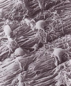
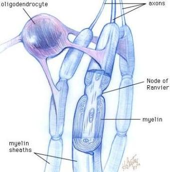
Oligodendrocytes are glial cells which form the myelin sheaths around axons in the central nervous system. These cells have a small number of cytoplasmic processes – the name oligodendrocyte comes from the Greek roots oligo, meaning ‘few’, dendro meaning ‘branch’, and kytos which denotes ‘cell’ – each of which forms a broad, flat structure after emanating from the cell body. An individual oligodendrocyte straddles a bundle of axons, and each of its processes wraps itself around the axon of a different neuron in the bundle. The processes form concentric circles of fatty tissue that make up a segment of that neuron’s myelin sheath; in between each segment of myelin is an unmyelinated section of the axon called the Node of Ranvier.
Myelin has a composition of 80% lipids and 20% protein, and performs a number of crucial functions in the nervous system. It insulates nerve fibres by reducing the leakage of ions through the cell membrane, and increases the velocity at which action potentials are conducted along the nerve fibre, by enabling them to ‘jump’ from one unmyelinated region to the next, in a process called saltatory conduction. Polio, multiple sclerosis, and other demyelinating diseases are caused by the breakdown of the myelin sheath in various regions of the nervous system, and can result in blindness and paralysis.
Oligodendrocyte progenitor cells (OPCs) are generated in the ventricular zone of the neural tube, in response to induction signals mediated by Sonic Hedgehog and other molecules. From the ventricular zone, the cells migrate outwards along the circumference of the tube, and then along its length. During this migration, OPCs actively seek axons around which they can wrap their processes once they have differentiated into oligodendrocytes. Myelination, the process by which oligodendrocytes wrap their processes around nerve fibres, begins towards the end of embryonic development, and continues postnatally.
A research team led by Bruce Appel, an associate professor of biology at Vanderbilt University Medical Center in Nashville, Tennessee, has produced time lapse movies of the migration of oligodendrocyte progenitor cells (OPCs) during zebrafish embryogenesis. The experiments, described in an advance online publication in Nature Neuroscience
Zebrafish embryos are transparent and are therefore one of the models of choice for developmental biologists. Appel’s team exploited this property of the embryos to make their movies. They first created transgenic zebrafish in which the OPCs express green fluorescent protein. Using a confocal microscope, they took large numbers of images of the cells, over a period of up to several days, as they migrated through the spinal cord. The images were then edited to produce time-lapse movies of the migrations.
The movies produced show that the process of OPC migration is far more dynamic than was previously thought. Throughout their migration, the cells extend and retract filopodia-like processes to obtain cues from their surroundings. Upon coming into contact with neighbouring OPCs, the processes are withdrawn and then extended in the opposite direction. This seems to be a mutual repulsion mechanism which ensures that OPCs are evenly distributed along the length of the axons they will myelinate.
The clip below shows the ameboid movement of OPCs as they migrate within the zebrafish spinal cord:
Appel and his colleagues then destroyed some of the existing OPCs by exposing them to laser light. They observed that cells which are migrating past the ablation site stop at the gaps and re-enter the cell cycle, thus generating new cells which then migrate into and fill the gaps:
“Now that we have a better understanding of OPC and oligodendrocyte behaviors, we are in a much better position to identify and study the genes that are necessary for myelination,” says Appel, “and having these genes in hand should aid in the design of drugs to promote remyelination following disease or injury.”
Update: Myelin to blame for many neuropsychiatric disorders
[According to]…UCLA neurology professor George Bartzokis…the miles of myelin coating in our brain are the key “evolutionary change that defines our uniqueness as a species” and, further, may also be the cause of “our unique vulnerability to highly prevalent neuropsychiatric disorders.” Bartzokis studied the reported effects of cholinergic treatments…[and] argues that [they] may have nonsynaptic effects as well, perhaps by enhancing myelination and myelin repair.
(via Biocompare Neuroscience News)


Those are some wicked videos!
I shudder to think how many PhDs and post-docs have been spent getting this to work. People have been going at this for at least a decade, and before that it was old-fashioned staining of fixed tissues. It’s cool, but a lot of work just for cool.
My son contracted Guillain-Barre (excuse the lack of accents) over ten years ago and, following three weeks of hospitalization and several months of rehab, was back playing competitive hockey. He was eight years old. While we, his parents, went through the ringer during the first months of his recovery and still are amazed at how well he recovered, the whole event is an insignificant part of his memory. Reading this article and watching these videos should give hope to those unfortunate enough to not have responded as completely to the body’s attempt at recovery from demyelinating conditions. Some of the research should focus on the differences in the recovery process (remyelination) between the pre-pubescent human and adults. Can anyone imagine what strides could be made in science if the same energy we put toward destroying each other could be put toward scientific research?
Pingback: Cells Weekly #5 « Migrations
Unbelievable
Pingback: It’s neurotransmission, but not as we know it « Neurophilosophy
Please note someone has inserted the words Sonic Hedgehog into the article.
to induction signals mediated by Sonic Hedgehog and other molecules. From the ventricular zone, the cells migrate outwards along the